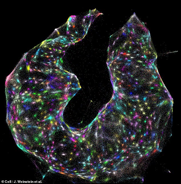
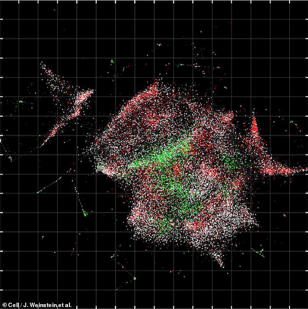
From readings of the complex interactions of these labels with the molecules and each other, a computer algorithm can work backward to reveal an image of the cell.
DNA microscopy could find myriad applications — including helping scientists study immune cells and tumours to develop new treatments to fight cancer.
The unorthodox imaging technique was developed by biophysicist Joshua Weinstein and colleagues at the Broad Institute in Cambridge, Massachusetts.
Unlike a traditional microscope that uses light to create an image, the new microscopy technique instead uses 'bar codes' of DNA that work to pinpoint the relative positions of molecules within a sample.
The approach can let scientists both assemble a picture of the cells they are studying while simultaneously revealing the cells genetic sequences.
'This gives us another layer of biology that we haven't been able to see,' said Dr Weinstein.
'It's an entirely new category of microscopy,' said paper co-author and computational biologist Aviv Regev.
'It's not just a new technique, it's a way of doing things that we haven't ever considered doing before,' she added.
The new imaging technique works by fixing cells into position in a reaction chamber and adding to them an assortment of DNA 'bar codes'.
These customised sequence of DNA work by sticking to the DNA and RNA molecules in the cells, giving each molecule a unique tag.
Once the tags latch on, they create more and more copies of each of the tagged molecules — hundreds in total — forming a growing pile which expands out from the original molecule's starting position.
'Picture every single molecule as a radio tower broadcasting its own signal outward,' Dr Weinstein said.
As the tagged molecule copies spread out, they eventually collide with other copies of tagged molecules — forcing them to link together to form distinctive paired genetic sequences that can be decoded using a DNA sequencer.
The closer the original molecules were to each other, the more likely their respective copies are to collide, resulting in more of their copies pairing.
In contrast, if the original molecules are further apart, then they ultimately produce less of the paired copies.
By analysing these paired sequences, a computer algorithm can work backwards to determine the location of each original molecule in the cells.

Remarkable footage shows how a revolutionary technique is letting scientists peer into cells at the genetic level — both imaging the cell and sequencing its DNA
The DNA-sequencing process can take up to 30 hours to process a sample, producing around 50 million DNA letters in genetic sequence for the computer to translate into both an image and a sequence of the cell's genome.
Because DNA can also attach to other molecules in cells beside DNA and RNA, the approach can also image and identify other cellular components including antibodies, receptors and even the molecules on tumours that immune cells target.
'You're basically able to reconstruct exactly what you see under a light microscope,' said Dr Weinstein.
In fact, he added, the two methods can be used to complement each other.
Whereas light-based microscopy can see molecules well even where they're present in limited numbers, DNA microscopy works well when molecules are present in dense concentrations.
The new technique can even handle the imaging of molecules that are piled up on top of each other.
The strength of the DNA microscopy technique lies in how it can combine the kinds of information produced by the two existing types of microscopy.
Basic optical microscopes, first invented in the early 1600s, use light to illuminate samples — and have been adapted in various ways that replace visible light with other wavelengths or electrons, for example or use samples that themselves glow.
Either way, all optical microscopes work on the principle that the sample gives off photons or electrons which can then be detected.
The second type of microscope relies on dissecting samples at a series of predefined locations and then combing this information to form a complete image.
While optical imaging can offer detailed portrayals of the structure and actions with a cell, dissection-style approaches can offer localised information like genetic data.
DNA microscopy, however, does both — capturing an picture of the cell's insides while also reading off the genetic sequences underpinning the image.
Concentrating on only one of these aspects, said Dr Weinstein, means that 'you're only getting part of the picture'.

From readings of the complex interactions of these labels with the molecules and each other, a computer algorithm can work backward to reveal an image of the cell
One potential application for DNA microscopy would be in accelerating the development of immunotherapy treatments that help patients to fight cancer.
Scientists might use the imaging method to identify the immune cells best suited for targeting a particular cancerous cell.
This could be achieved because every cell has a unique DNA coding, or genotype, that results in given observable characteristics — its so-called phenotype — depending on the environment in which the cell develops.
'By capturing information directly from the molecules being studied, DNA microscopy opens up a new way of connecting genotype to phenotype,' said paper author and biochemist Feng Zhang.
According to Weinstein, DNA microscopy is especially well suited to studying immune cells, in which both even single-letter DNA changes and a the cell's location within surrounding tissue can radically alter the antibodies the cell produces.
However, the possibilities for new research using this technique are broad and rich for exploration, Professor Regev said.
'We hope that it sparks the imagination — that people will be inspired with great ideas that we've never thought about,' she added.
The Daily Mail
More about: DNA


